Paris Museum
- Details
- Category: Paris Museum
- Hits: 729
Holothuria flammea Quoy & Gaimard, 1833: 117, pl. 6, figs 5-6.
Stichopus flammeus; Brandt, 1835: 73; Selenka, 1867: 320.
Type data: EcHh 3270; Vanikoro (Phlippines), unknown depth; coll. Quoy & Gaimard; 1829; well preserved; well-relaxed; ventro-longitudinal dissection; specimen partly eviscerated.
Anatomical description: 65 mm long; 15-25 mm wide; bivium arched; trivium flattened; mouth ventral; anus terminal; dorsal body wall beige brown, mottled with darker brown; ventral body wall beige-brown; tentacles beige; dorsal appendages beige-brown; ventral tube feet beige; dorsal papillae in distinct rows along the ambulacrae; ventral tube feet dispersed all over trivium, but mainly in ambulacrae; bivium and trivium not separated by lateral fringe of appendages; body wall 6-8 mm thick, rough to the touch; 15 small tentacles counted; radial plates with straight posterior side; radial plates twice as wide as interradial ones; radial plates 3 times longer than interradial ones; number of tentacle ampullae could not be determined; tentacle ampullae 4 mm long; number of Polian vesicles could not be determined; presence of stone canals could not be determined; gonad not observed; longitudinal muscles bifid, very wide, touching each other, attached at edges. Cuvierian tubules absent.
Ossicle description: tentacles with non-bifuracating rods of various sizes, slightly rugose distally; dorsal and ventral body wall witrh tables with roundish disc perforated by four central holes and one ring of peripheral holes, spire with one cross-beam, ending in a narrow simple crown, buttons regular, with smooth rim, and generally perforated by three pairs of holes; dorsal appendages with tables similar to those of the body wall and with buttons with 3-5 pairs of holes and with rod-like buttons; ventral tube feet with tables and buttons silmilar to those of the body wall and with in addition also perforated plates; longitudinal muscles with O-shaped ossicles; cloaca with smooth irregular rods; loacal retractor muscles and respiratory tree devoid of ossicles; ossicle assemblage of the gonad, rete mirabile and gut was not determined.
Known distribution: Vanikoro, Philippines.
Taxonomic decision: junior subjective synonym of H. monacaria Lesson, 1830 (taxonomic decision for synonymy based upon re-examination of holotype of H. flammea and description of H. monacaria) of which no more type material exists. The name H. monacaria is to be stabilized by the designation of a neotype.
Remarks: Holothuria flammea is redescribed by Cherbonnier (1952) whereby he notes that the species is, without doubt, a synonym of Holothuria monacaria Lesson, 1830 (see also the original label on the jar; the name Stichopus monacaria Lesson can still be read). H. monacaria, in turn, is generally regarded as one of the many subjective synonyms of H. hilla[1]. The ossicle assemblage of H. flammea indeed indicates that H. flammea, just as H. hilla, belongs to Mertensiothuria. However, it is unlikely that H. flammea is also a synonym of H. hilla (see also the redescription of H. hilla) given that: (i) H. flammea does not present Cuvierian tubules whereas H. hilla generally is considered to possess these (see also remarks with H. hilla) and (iii) the ossicles of the cloaca are irregular smooth slender rods in H. flammea whereas they are spiky rods in H. hilla.
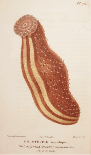
Figure – Original drawing of Holothuria monacaria Lesson, 1830.
For original description click here.
{morfeo 35}
[1] Based upon non-type material also collected by Lesson & Garnot in Borabora, Cherbonnier (1951) suggested that H. monacaria might be a synonym of A. mauritiana. This is very doubtful as Lesson (1830) clearly stated that H. monacaria has 16-20 tentacles and that the anus is devoid of teeth.
- Details
- Category: Paris Museum
- Hits: 855
Holothuria edulis Lesson, 1830: 125, pl. 46, fig. 2; Selenka, 1867: 341; Semper, 1868: 89, 278, pl. 31, fig. 7, pl. 32, fig. 4, pl. 33, fig. 3, pl. 36, figs 2, 5, 9, 10; Semper, 1869: 120; Ludwig, 1882: 137; Lampert, 1885: 81; Théel, 1886: 216; Ludwig, 1887: 1227; Ludwig, 1888: 807; Ludwig, 1889-92: 329; Saville-Kent, 1893: 233, 237; Sluiter, 1894: 103; Koehler, 1895: 281; Sluiter, 1895: 79 ; Bedford, 1898: 147; Ludwig, 1899: 559; Sluiter, 1901: 8; Koningsberger, 1904: 49; Koehler & Vaney, 1908: 7; Bedot, 1909: 60; Mitsukuri, 1912: 77; Pearson, 1913: 69, pl. 9, fig. 2; Erwe, 1913: 183; Clark, 1921: 177, pl. 19, fig. 1; Clark, 1923: 421; Clark, 1925: 103; Panning, 1928: 231, figs 35, 36; Schmidt, 1930: 465; Clark, 1932: 231 ; Domantay, 1936: 12, pl. 1, figs. 1-2; Pabish, 1936:101; Pabish, 1936: 2185 ; Clark, 1938: 519 ; Sella & Sella, 1940: 14, 39 ; Clark, 1946: 427 ; Dawydoff, 1952: 117; Endean, 1953: 56 ; Domantay, 1954: 342 ; Endean, 1956: 131 ; Endean, 1957: 253 ; Domantay, 1960: 87, fig. 6a-c; Domantay, 1962: 87, fig. 6a-c; Macnae & Kalk, 1962: 108 ; James, 1969: 61 ; Townsley & Townsley, 1972: 176; Daniel & Halder, 1974 : 428 ; Waren, 1980 : 295 ; Grosenbaugh, 1981 : 51 ; James, 1982 : 5 ; Tortonese, 1977 : 275 ; James, 1983 : 98; Brouns & Heijs, 1985: 175; Levin & Sayapina, 1988; 122; James, 1988: 404 Allen & Steene, 1994: 244 (colour plate) ; Fiege et al., 1994: 86; Klinger et al., 1994: 524ss; Colin & Arneson, 1995: 260, fig. 1231 (colour plate); Weinberg, 1997: 245 (colour plate); Klinger & Johnson, 1998: 467ss; Baine & Forbes, 1998: 4 (colour plate); Debelius, 1998: 300 (colour plate); Liao, 1998: 80; Uthicke, 1998: 532ss; Zulfigar & Tan Shau Whai, 1999: 76 ; Roberts et al., 2000: 264, fig. 3b; Marshall et al. 2001: 46, 47, 54; Zulfigar et al., 2001: 364; James, 2001: 7, fig. 13 (B/W photo); Skewes et al., 2002: 13; Chen, 2003: 20; Lane, 2004: 232; Rasolofonirina et al., 2004: 137; Skewes et al., 2004: appendix 3-4; Mmbaga & Mgaya, 2006: 3ss.
Holothuria (Holothuria) edulis; Panning, 1935: 43,fig. 36a-d; Domantay, 1936: 398; Van den Spiegel & Jangoux, 1989: 225.
Halodeima edulis; Pearson, 1913: 69, pl. 9, fig. 12; Ohshima, 1935: 144; Heding, 1939: 220; Panning, 1944: 65, fig. 32; Cherbonnier, 1951: 399, fig. 3; Cherbonnier, 1951: 81; Clark, 1952: 204 ; Cherbonnier, 1955: 142, pl. 29, fig. c; Cherbonnier, 1959: 250; Cherbonnier, 1963: 5 ; Chang & Liao, 1963: 67, fig. 12, pls 2, 6; Chang & Liao, 1964: 35, fig. a-c; Cherbonnier, 1967: 56; Cherbonnier, 1979: 861; Mergner, 1979: 481; Intes & Ménou, 1979: 12, 20 (map), 23 (BW plate).
Holothuria (Halodeima) edulis; Rowe, 1969: 138; Clark & Rowe, 1971: 176, pl. 27, fig. 14; Rowe & Doty, 1977: 231, figs 3a, 7b; Levin, 1979: 20; Levin, 1979b: 22; Roberts, 1979: 45ss; Cherbonnier, 1980: 632, fig. 9A-L; Levin, 1980: 52; Liao, 1980: 115; Mary Bai, 1980: 12, text fig. 9E; Humes, 1980: 72, 74, 86, 87; Grosenbaugh, 1981: 51; Price, 1981: 9; Price, 1982: 11; Hoskin & Waren, 1983: 24; James, 1983: 100; Price, 1983: 87, 91, fig. 48; Rowe, 1983: 156; Soota et al., 1983 : 519; Liao, 1984: 222; Thandar, 1984: 276; Reyes-Leonardo, 1984: 146, pl. 3, fgi. 1A-I; Conand & Chardy, 1985: 295; Reyes-Leonardo et al., 1985: 272; James, 1985[1988]: 404; Price & Reid, 1985: 4; Cherbonnier, 1986: 43; James, 1986: 585 ; Marsh, 1986: 73 ;Cannon & Silver, 1986: 22, fig. 6f, text fig.; Cherbonnier & Féral, 1986: 87 (colour plate) ; George & George, 1987 : 246 ; Mokhopadhyay, 1988 : 6, fig. 4a-b ; Cherbonnier, 1988 : 75, fig. 29A-I ; Conand, 1989 : 23, fig. 2 ; Chambers, 1989: 89 ; Jangoux et al., 1989 : 163; Kalashnikof, 1989: 64, fig. 2; Levin & Dao Tan Ho, 1989: 55; Féral, 1990: 367, fig. 279 (colour photo); Marsh et al, 1993: 64; Marsh, 1994: 10; Marsh, 1994: 57; Holland, 1994: 2; Kerr, 1994: 168; Sant, 1995: 27; James, 1995: 52, fig. 1F-G; Pawson, 1995: 188; Rowe & Gates, 1995: 291; Liao & Clark, 1995: 436, fig.252a-c; Gosliner et al., 1996: 279, fig. 1029 (colour plate); Massin, 1996: 19, fgi. 11A-G; Tsuda, 1997: 16 ; Liao, 1997; 101, fig. 56a-d; Rowe & Richmond, 1997: 304 (colour drawing); Conand, 1998: 1177, text fig.; Massin, 1999: 21, figs 14 (map), 110d (colour plate); Bussarawit & Thongtham, 1999: 35; Uthicke & Karez, 1999: 71ss; Forbes et al., 1999: 30 (colour plate); Schoppe, 2000: 111 (colour plate); Samyn, 2000 : 15; Lane et al. 2000: 488 ; Samyn &Vanden Berghe, 2000: 5, 17, 22; Desurmont, 2003: 10; Samyn, 2003 : 36, fig. 15A-E ; Paulay, 2003: 577; Putchakarn & Sonchaeng, 2004: 425; Marsh & Morrison, 2004: 303, 339; Rowe & Richmond, 2004: 3301; Thandar & Samyn, 2004 : 255; Kumara et al., 2005: 25; Sastry, 2005: 105; O’Loughlin et al., 2007: 43, fig. 5c-f; 45(color pictures): fig 45 (ossicle assemblage).
Trepang edulis; Jaeger, 1833: 24; Brandt, 1835: 57.
Holothuria fuscocinerea; Selenka, 1867: 337, pl. 19, fig. 86 (non H. fuscocinerea Jaeger, 1833); Poscidia, 1968: xxx ; Verbist, 1993: 116; Chen, 2003: 20.
Type data: EcHh 543; Moluccas, (Indonesia); unknonwn depth; coll. M.M. Lesson & Garnot during Expedition Duperrey; 1825; well preserved; well relaxed; ventro-longitudinal dissection.
Anatomical description: 160 mm long; 19-24 mm wide; bivium arched; trivium flattened; mouth ventral; anus terminal; dorsal body wall white grey-brown; ventral body wall grey-beige; tentacles light beige; dorsal appendages brownish; ventral tube feet brownish; dorsal appendages dispersed regularly all over bivium; ventral tube feet dispersed regularly all over trivium; bivium and trivium not separated by lateral fringe of appendages; body wall ±2 mm thick, smooth to the touch; 18 tentacles counted; structure of the calcareous ring was not assessed; number of tentacle ampullae could not be determined; number of Polian vesicles could not be determined; presence of stone canals could not be determined; gonad not observed; longitudinal muscles single, wide, attached at edges. Presence of Cuvierian tubules could not be assessed.
Ossicle description: tentacles with spiny rods of various sizes, large ones perforated distally; dorsal and ventral body wall with tables and button-like rosettes, tables with disc smooth, reduced, with four pillars united by a single cross beam and ending in a crown in the form of a Maltese crown; dorsal appendages with tables and rosettes similar to those of body wall and with rods; ventral tube feet with tables and rosettes as in body wall and in addition elongated button-like rosettes; longitudinal and cloacal retractor muscles, cloaca and respiratory tree devoid of ossicles; ossicle assemblage of gonad, rete mirabile and gut was not assessed.
Known distribution: Red Sea (Gulf of Suez, Shab Mahmoud, Gulf of Aqaba, Eilat, Dahab, Abu Zabad, Eritrea Entedebir), Kenya, Aden, Zanzibar, Mozambique, Tanzania, Madagascar, Oman (Muscat), Persian Gulf, Maldive Islands, Sri Lanka, India (Laccadive Islands, Gulf of Mannar, Andaman Islands, Nicobar Islands), Cocos Keeling Islands, Indonesia (Sumatra, Java, Makassar Strait, Sulawesi, Salayer, Komodo, Timor, Ceram, Pari Islands Ambon, Moluccas = type locality, Irian Jaya), Malaysia (Mainland, Shaba), Australia (GBR, NE coast, Tasman Sea, NW coast, QLD, NSW, WA, Dampier Archipelago; NT), Philippines (Bohol), Thailand, Vietnam, China, Japan, Mariana Islands (Guam, Saipan), Caroline Island (Chuuk (= Truck) Atoll, Tonoas Island, Yap), Papua New Guinea (Port Moresby, New Britain), Solomon Islands, New Caledonia, Loyalty Islands (Lifu), Vanuatu (= New Hebrides), Fiji, Line Islands (Fanning Island), Society Islands (Tahiti), Pitcairn Island. (H. signata records still need to be taken out, also from citation list)
Taxonomic decision: valid species (confirmed after re-examination of the lectotype and other voucher specimens).
For original description click here.
{morfeo 34}
- Details
- Category: Paris Museum
- Hits: 607
Holothuria (Cystipus) dura Cherbonnier & Féral, 1981: 385, fig. 17A-M; Samyn, 2003: 30; Laguerda-Figueras & Solis-Marin, 2009: 577.
Type data: EcHh 3000; Station 12: 14°00,8 N-120°20,5 E (Philippines); 210-187 m depth; coll. Forest J. (Mission MUSOROSTOM); 20.III.1976; well preserved; poorly relaxed; ventro-longitudinal dissection; calcareous ring and associated structures have been removed; specimen largely eviscerated.
Anatomical description: 120 mm long; 18-21 mm wide; bivium arched; trivium not distinctly flattened; mouth ventral; anus terminal; dorsal body wall white to light beige with brown patches; ventral body wall white to light brown; tentacles white to light beige; dorsal appendages white; ventral tube feet beige; dorsal appendages few, arrangement could not be determined; ventral tube feet few, arrangement could not be determined; bivium and trivium not separated by lateral fringe of appendages; body wall ±2 mm thick, rough to the touch; 15 tentacles counted; radial plates with a concave posterior side, interradial plates narrower than radial ones, radial plates approximately two times longer than interradial ones; 17 tentacle ampullae counted; number of Polian vesicles could not be determined; presence of stone canals could not be determined; gonad reduced, with unbranching tubules; longitudinal muscles bifid, wide, free at edges. Presence of Cuvierian tubules could not be assessed.
Ossicle description: tentacles with rods of various sizes, rugose, distally branching or perforated; dorsal body wall with few tables with undulating spiny and roundish disc perforated by one central hole and 1-2 rings of peripheral holes, four short pillars united by one cross beam ending in a narrow crown and abundant rugose buttons, often twisted given the appearance of a fenestrated ellipsoid, with undulating rim and with 4-6 pairs of holes; ventral body wall with buttons only, stouter but smaller and less twisted than those of the dorsal body wall, perforated with 3 to occassionally 4 pairs of holes; dorsal appendages with tables similar to those of body wall, buttons much less rugose and not twisted as those of body wall and perforated rod-like plates of various length;ventral tube feet with very few tables simlilar to those of body wall and numberous buttons with up to nine pairs of holes; anal papillae with ossicle assemblage similar to that of dorsal appendages; longitudinal and cloacal retractor muscles, gonad, respiratory tree and cloaca devoid of ossicles. Ossicle assemblage of Rete mirabile and gut was not assessed.
Known distribution: 14°00,8 N-120°20,5 E (Philippines).
Taxonomic decision: valid species (confirmed after re-examination of the holotype).
For original description click here.
{morfeo 33}
- Details
- Category: Paris Museum
- Hits: 631
Holothuria (Lessonothuria) duoturricula Cherbonnier, 1988: 119, fig. 48A-N; Samyn, 2003: 39.
Type data: EcHh 2655; N°AA-1, Ambatoloaka Beach, Nosi Be (Madagascar); collected in the intertidal; coll. G. Cherbonnier; 27.V.1960; well preserved; poorly relaxed; dissected ventro-longitudinally; calcareous ring was separated from the body.
Anatomical description: 50 mm long; 7-16 mm wide; bivium arched; trivium flattened; mouth ventral; anus terminal; dorsal body wall brown, mottled with beige; ventral body wall brown; tentacles brown; dorsal appendages beige; ventral tube feet light brown; dorsal appendages spread regularly over complete bivium; ventral tube feet in indistinct rows along the ambulacrae; bivium and trivium not separated by lateral fringe of appendages; body wall ±1 mm thick, rough to the touch; 23 tentacles counted; radial plates with a slightly indented posterior side, interradial plates narrower than radial ones, radial plates approximately two times longer than interradial ones; number of tentacle ampullae could not be determined; number of Polian vesicles could not be determined; presence of stone canals could not be determined; gonad not observed; longitudinal muscles bifid, very wide, flat, attached at edges. Cuvierian tubules absent.
Ossicle description: tentacles with rods, swollen at ends; dorsal body wall with tables and buttons, table disc perforated by a central hole and one circle of peripheral perforations, rim spiny, spire with short pillars with 0-1 crossbeams ending in a open spiny crown; buttons irregular,, twisted, with 4-8 holes; ventral tube feet with tables similar to those of body wall, regular buttons with 4-6 pairs of holes and perforated plates with up) to 6 rows of holes; longitudinal muscles, gut and cloaca devoid of ossicles; ossicle assemblage of dorsal appendages, cloacal retractor muscles, gonad, respiratory tree and rete mirabile could not be determined.
Known distribution: Nosi-Bé, Madagascar.
Taxonomic decision: valid species (confirmed after re-examination of the holotype).
For original description click here.
{morfeo 32}
Page 4 of 6