Paris Museum
- Details
- Category: Paris Museum
- Hits: 644
Holothuria jousseaumei Cherbonnier, 1954: 255; Cherbonnier, 1955: 147, pl. 34, fig. a-p ; Cherbonnier, 1967: 57, 61 ; Daniel & Halder, 1974: 417; Tortonese, 1977: 275.
Holothuria (Cystipus) jousseaumei; Rowe, 1969: 156; Clark& Rowe, 1971:176; Samyn, 2003: 30, 35, 121.
Type data: EcHh 582; Red Sea; depth not provided; coll. M. Botta, 1837; well preserved; well relaxed; ventro-longitudinal dissection with anal side severely damaged.
Anatomical description: 58 mm long; 12-20 mm wide; bivium arched; trivium flattened; mouth ventral; anus terminal; dorsal body wall light beige; ventral body wall light beige; tentacle colour brownish; dorsal appendages light beige; ventral tube feet light beige; dorsal appendages dispersed all over bivium but with one clear row along lateral side of ambulacrum; ventral tube feet in distinct rows; bivium and trivium not separated by a lateral fringe of appendages; body wall 1 mm thick, rough to the touch; tentacles small, peltate with shallow indentions, number could not be determined; radial plates with slightly convex posterior side, interradial plates only slightly narrower than radial ones; radial plates 1,5 x as long as interradial ones; number of tentacle ampullae could not be determined; number of Polian vesicles could not be determined; number of stone canals could not be determined; gonad in single tuft with ramified, well-developed long brown tubes; longitudinal muscles narrow, free at edges; Cuvierian tubules not observed.
Ossicle description: tentacles with few slender, smooth rods; dorsal and ventral body with tables and buttons, tables with very low spire ending in a very nodulous crown giving a morula-like impression or with a more narrow crown, buttons with 2-8 pairs of perforations (generally with 4 ), very nodulous both marginally and medianly; dorsal appendages with reduced tables, elongated smooth buttons with up to 10 pairs of perforations, rods widened and perforated distally and centrally, and perforated plates; ventral tube feet with ossicle assemblage as in dorsal assemblage , but without perforated plates; longitudinal and cloacal retractor muscles, gut, respiratory tree, cloaca and gonad devoid of ossicles;ossicle assemblage of rete mirabile was not assessed.
Known distribution: Red Sea (Djibouti, Entedebir, Gulf of Aqaba), Seychelles (Mahé).
Taxonomic decision: valid species (confirmed after examination of the type series).
For original description click here.
{morfeo 46}
- Details
- Category: Paris Museum
- Hits: 640
Holothura (Platyperona) insolita Cherbonnier, 1988: 101, fig. 41A-M; Samyn, 2003: 65.
Type data: EcHh 3548; Tuléar, Station 450 (Madagascar); depth not provided; coll. Thomassin, date not given; well preserved; poorly relaxed; ventro-longitudinal dissection; specimen eviscerated; generally in poor shape.
Anatomical description: 24 mm long; 7-10 mm wide; bivium arched; trivium flattened; mouth ventral; position of anus could not be determined; dorsal body wall grey beige; ventral body wall white to beige with transversal narrow brown bands anteriorly and minutes spots over the rest of the body; tentacle colour yellow-brown; dorsal appendages white; ventral tube feet white; dorsal appendages few, dispersed all over bivium; ventral tube feet few, dispersed over complete trivium; bivium and trivium not separated by a lateral fringe of appendages; body wall <1 mm thick, rough to the touch; tentacles small, number could not be determined; radial plates with concave posterior side, interradial plates as wide as radial ones; radial plates 1,5-2 x as long as interradial ones; number of tentacle ampullae could not be determined; single Polian vesicles observed; single contorted, stone canal observed; gonad was not observed; longitudinal muscles narrow, largely transparent, free at edges; Cuvierian tubules not observed.
Ossicle description: tentacles with few slender unbranched rods; dorsal and ventral body wall with tables with round disc perforated by one central hole and one ring of peripheral holes, with spire united by 1 to 2 crossbeams ending in a narrow crown, dorsal appendages with tables similar to those of the body wall, but with 1-3 cross-beams, and rods; ventral tube feet with tables similar to those of the body wall and with rod-like buttons; longitudinal muscles, respiratory tree and gut devoid of ossicles; ossicle assembalge of cloacal retractor muscles, gonad, cloaca and rete mirabile were not assessed.
Known distribution: only known from the type locality.
Taxonomic decision: nomen inquirendum (to be compared with Mesothuria spp.).
Remarks: Cherbonnier (1988) mentions that he found buttons in the anal region. Given the small size of the specimen, we however did not take tissue from that region. We can thus not confirm Cherbonnier’s observation. Actually, it is our impression that this species possibly does not belong to the genus Holothuria.
For original description click here.
{morfeo 45}
- Details
- Category: Paris Museum
- Hits: 674
Holothuria (Fistularia) hilla Lesson, 1830: 226.
Holothuria (Fistularia) Lilla; Lesson, 1830: 227, pl. 78. (lapsus calami).
Holothuria hilla; Cherbonnier, 1951: 532, fig. 1; Tortonese, 1953: 42, fig. 5; Cherbonnier, 1955: 153, pl. 32, fig. g-r; Macnae & Kalk, 1958: 36ss; Kalk, 1958: 213ss; Kalk, 1959: 7, 22; Macnae & Kalk, 1962 104ss; James, 1969: 62; Cherbonnier, 1963: 5; Cherbonnier, 1966: 56; Nagabhushanam & Rao, 1972: 290; Lawrence, 1980: 202; Grosenbaugh, 1981: 51; Branch & Branch, 1981: 249; Kropp, 1982: 446, 449; James, 1983: 98; James, 1983: 93; James, 1988: 404; Zoutendijk, 1989: 2; Colin & Arneson, 1995: 262, fig. 1234 (colour plate); James, 1995: 273; Weinberg, 1997: 246 (colour plate); Solis-Marin et al., 1997: 256; Hickman, 1998: 47 (colour plate); Lioa, 1998: 80; Kerr et al., 1998: 786; Conand, 1999: 10ss; Baine & Forbes, 1998: 4; Zulfigar & Tan Shau Hwai, 1999: 76; Roberts et al., 2000: 264, fig. 3d; James 2001: 7, fig. 15, (B/W photo); Zulfigar et al., 2001: 364; Conand & Mangion 2002: 28.
Holothuria (Holothuria) hilla; Vandenspiegel & Jangoux, 1989: 225.
Holothuria (Thymiosycia) hilla; Rowe, 1969: 147; Clark & Rowe, 1971: 178, pl. 28, fig. 9; A.M. Clark & Taylor, 1971: 91; Liao, 1975: 214; Rowe & Doty, 1977: 232, figs 4b, 8b; Levin, 1979: 22; Sloan et al., 1979: 123; Levin, 1980: 53; Liao, 1980, 115; Mary Bai, 1980: 13, textfig. 9I; Tortonese, 1980: 107; Humphreys, 1981: 35; Price, 1981: 9; Price, 1982: 11; fig. 51a-d'; Mukhopadhyay & Samanta, 1983: 307, fig. 8A-C; Price, 1983: 93; Rowe, 1983: 158; Leonardo & Cowan, 1984: 38, textfig; .Reyes-Leonardo, 1984a: 147, pl. 4 fig. 2a-f; Liao, 1984: 222; A.M. Clark, 1984: 99; Conand & Chaudry, 1985: 295; Richard, 1985: 457; James, 1985 [1988]: 404; Price & Reid, 1985: 6; Marsh, 1986: 73; Cannon & Silver, 1986: 25, fig. 7e, textfig.; Féral & Cherbonnier, 1986: 92 (colour plate); Cutress & Rowe, 1987: 267, figs 2c, 6e; George & George, 1987: 247; Cherbonnier, 1988: 85, fig. 34A-L; Mukhopadhyay, 1988: 8, fig. 7a-b1; Jangoux et al., 1989: 163; Conand, 1989: 28; Chao & Chang, 1989: 118, figs 17, 30D; Pauley, 1989: 27; James, 1989: 126; Levin & Dao Tan Ho, 1989: 57; Imaoka, 1991: 178, fig. 3A-D; James, 1991: 23; Mukhopadhyay, 1991: 407; Kerr et al., 1993: 782ss; Marsh et al., 1993: 64; Kerr, 1994: 169; Marsh, 1994a: 11; Marsh, 1994b: 57; Rowe & Gates, 1995: 302; Liao & A.M. Clark, 1995: 463, fig. 276a-d; James, 1995a: 59, pl. 1D, fig. 2G-H; Pawson, 1995: 189; Massin, 1996b: 30, fig. 20A-G; Gosliner et al., 1996: 280, fig. 1032 (colour plate); Liao, 1997: 141, fig. 83a-d; Rowe & Richmond, 1997: 304 (colour drawing); Liao, 1998: 80; Erhardt & Beansch, 1998: 1084 (colour plate); Forbes et al., 1999; 42, textfig + colour plate + map; Bussarawit & Thongtham, 1999: 35 ; Massin, 1999: 55 figs 44 (map), 11D (colour plate); Samyn, 2000: 15; Lane et al., 2000: 489; Samyn & Vanden Berghe, 2000: 28; Schoppe, 2000: 113, colour plate; Putchakarn & Sonchaeng, 2004: 426; Sastry et al., 2004: 64; James, 2004: 123; Marsh & Morrison, 204: 303, 339; Thandar & Samyn, 2004: 255: Kumara et al., 2005: 25; Solis-Marin et al., 2005: 133; Sastry, 2005: 110.
Holothuria (Mertensiothuria) hilla; Samyn & Massin, 2003: 2500, figs 5A-E, 11C, 12F (colour plate); Samyn, 2003: 45, fig. 53A (map); Rowe & Richmond, 2004: 3301; Samyn et al., 2005: 15.
Type data: EcHh 542; Borabora (Tahiti); depth not given; coll. Lesson & Garnot (Exp. Dupperey, 1825); well preserved; well relaxed; ventro-longitudinal dissection; calcareous ring damaged due to dissection; specimen partly eviscerated.
Anatomical description: 85 mm long; 10-15 mm wide; bivium arched; trivium not distinctively flattened; mouth ventral; anus terminal; dorsal body wall cream; ventral body wall cream; tentacle colour could not be determined; dorsal appendages cream; ventral tube feet cream; position of dorsal appendages could not be determined; position of ventral tube feet could not be determined; bivium and trivium not separated by a lateral fringe of appendages; body wall 1,5-3 mm thick, smooth to the touch; tentacles cut from the spcecimen, number could not be determined; structure of calcareous ring could not be adequately assessed, the radial plates with slightly indented posterior side; number of tentacle ampullae could not be determined; number of Polian vesicles could not be determined; number of stone canals could not be determined; gonad not observed; longitudinal muscles narrow and flat, bifid, free at edges; Cuvierian tubules not observed.
Ossicle description: dorsal and ventral body wall with tables and buttons, tables with smooth disc, perforated by four central holes and one ring of peripheral holes, spire low with one cross-beam, ending in a narrow crown, buttons with 3-4 pairs of holes; dorsal tube feet with tables and buttons similar to those of body wall and with rod-like buttons; ventral tube feet with tables and buttons as in body wall and with perforated plates; cloaca with spiky rods; longitudinal muscles with O-shaped deposits and buttons with two holes; cloacal retractor muscles devoid of ossicles; ossicle assemblage of the tentacles, gonad, respiratory tree, rete mirabile and gut was not assessed
Known distribution: cannot be assessed (see remarks).
Taxonomic decision: valid species (confirmed after examination of the holotype)
Remarks: The original illustration of H. hilla (cf. figure 2) does not fit with what current researchers (see for instance Erhardt & Baensch, 1998: 1084) call H. hilla. However, the ossicle assemblage fits neatly with the currently applied concept of H. hilla (see for instance Samyn et al, 2003 (p. 2501).
To establish the distribution pattern of H. hilla, all known records of should be evaluated against the original drawing of Lesson.
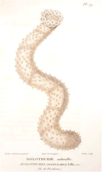
Fig. Original drawing of H. hilla; remark the very clearly drawn transversal banding, the many tube feet and the coloration pattern.
For original description click here.
{morfeo 44}
- Details
- Category: Paris Museum
- Hits: 653
Holothuria (Semperothuria) granosa Cherbonier, 1980: 66, fig. 25A-K; Samyn, 2003:77.
Type data: EcHh 2727; Station 1/16, Tuléar, Luce, North of Fort-Dauphin (Madagascar); intertidal; coll. A. Crosnier; 1960; well preserved; poorly relaxed; ventro-longitudinal dissection with anal side severely damaged; calcareous ring removed from specimen; specimen partly eviscerated.
Anatomical description: 82 mm long; ± 20 mm wide; bivium arched; trivium ot flattened; mouth terminal; anus terminal; dorsal body wall beige mottled with brown; ventral body wall beige-brown; tentacles black; dorsal appendages brown; ventral tube feet brown; dorsal appendages dispersed regularily over complete bivium; ventral tube feet dispersed regularily over complete trivium; bivium and trivium not separated by a lateral fringe of appendages; body wall 2 mm thick, smooth to the touch; tentacles rather small, 13 counted; radial plates with slightly indented posterior side; radial plates roughly 2 times wider than interradial ones; radial plates roughly of same length as interradial ones; number of tentacle ampullae could not be determined; three Polian vesicles, 12-19 mm long; number of stone canals could not be determined; gonad not observed; longitudinal muscles narrow and flat, bifid, attached at edges; Cuvierian tubules not observed.
Ossicle description: tentacles with nearly uniform sized rugose rods; dorsal body wall with tables with reduced smooth disc, high spire ending in a Maltese cross and rugose rods; ventral body wall with rugose rods only; longitudinal and cloacal retractor muscles, cloaca, respiratory tree and gut devoid of ossicles; ossicle assemblage of dorsal and ventral appendages and rete mirabile was not assessed.
Known distribution: île Sainte Luce (Madagascar).
Taxonomic decision: valid species (confirmed after re-examination of the holotype).
For original description click here.
{morfeo 43}
Page 3 of 6